Tendon Diagram - Tendons Basic Science Orthobullets. The anterior cruciate ligament prevents the femur from sliding backward on the tibia (or the tibia sliding forward on the femur). The fleshy, thick part of the muscle is called its belly. Cook and purdum have proposed a new strategy when approaching tendon pain, and this is called the tendon continuum. Allows the foot to be turned inward and also supports the arch of the foot. Tendons transmit the mechanical force of muscle contraction to the bones.
Tendon diagram you searching for is usable for you. The achilles tendon is the largest. A partial tear is when one of the tendons of the rotator cuff is frayed or damaged. Related posts of foot tendons and ligaments diagram ankle bones anatomy structure. In the leg muscles diagram above, there are many muscles that make up your legs and support it to move.
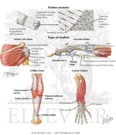
Tendon Anatomy from www.netterimages.com If you tear the biceps tendon at the shoulder, you may lose some strength in your arm and have pain when you forcefully turn your arm from palm down to palm up. The achilles tendon or heel cord, also known as the calcaneal tendon, is a tendon at the back of the lower leg, and is the thickest in the human body. It serves to attach the plantaris, gastrocnemius (calf) and soleus muscles to the calcaneus (heel) bone. The patellar tendon holds the patella with other two bones, similarly iliotibial band helps in supporting tibia and fibula. Related posts of foot tendons and ligaments diagram ankle bones anatomy structure. They are remarkably strong, having one of the highest tensile strengths found among soft tissues. Attaches the calf muscles to the calcaneus, most important muscles for running, jumping, walking etc. A muscle's origin is where a tendon attaches it to the *less* movable bone.
If you tear the biceps tendon at the shoulder, you may lose some strength in your arm and have pain when you forcefully turn your arm from palm down to palm up.
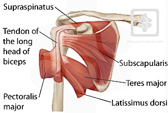
A tendon is a band of tissue that connects a muscle to a bone. Tendons in the knee play a very important role in holding the knee and the muscles together. The fleshy, thick part of the muscle is called its belly. The achilles tendon is a tough band of fibrous tissue that connects the calf muscles to the heel bone (calcaneus). One of the most important tendons in terms of mobility of the leg is the achilles tendon. Connect by text or video with a u.s. 2 ligaments (trapezoid& conoid ligaments) attach the clavicle coracoid process of scapula these tiny ligaments (w/ acominoclavicular joint) keep scapula attached to clavicle. Learn about the anatomy and physiology of tendons. It can be used by a teacher or student for academic purposes. Diagram of the shoulder, including the location of the rotator cuff. Attaches the calf muscles to the calcaneus, most important muscles for running, jumping, walking etc. Allows the action of raising the foot. Tendon, tissue that attaches a muscle to other body parts, usually bones.
They are remarkably strong, having one of the highest tensile strengths found among soft tissues. Diagram of knee tendons and ligaments. Anatomynote.com found right arm muscle and tendon anatomy from plenty of anatomical pictures on the internet. Start studying muscles and tendons. Reflex exam (deep tendon reflexes).
Shoulder Tendons Shoulderdoc from www.shoulderdoc.co.uk Tendons, located at each end of a muscle, attach muscle to bone. In the back and elsewhere in the body, tendons attach muscles to bones. If you would like to learn all the parts of the foot structure, you have come to the right place. Diagram of knee tendons and ligaments. They propose there are 3 stages to this continuum. Tendons in the knee play a very important role in holding the knee and the muscles together. Ankle bones anatomy structure 10 photos of the ankle bones anatomy structure ankle tendons anatomy, elbow bones anatomy, hand bones anatomy, leg bones anatomy, shoulder bones anatomy, tibia anatomy, wrist bones anatomy, foot, ankle tendons anatomy, elbow bones anatomy, hand bones anatomy, leg bones anatomy. Allows the action of raising the foot.
And vilensky j all in one anatomy exam review.
Fall on one point of shoulder and can rupture these ligaments with dislocation of ac joint. Ligaments join the knee bones and provide stability to the knee: This important tendon in the back of the calf and ankle stores the elastic energy needed for running, jumping, and other physical activity. This forearm muscle is responsible for extending all of the fingers of the hand except the thumb. Tendons in the knee play a very important role in holding the knee and the muscles together. In the leg muscles diagram above, there are many muscles that make up your legs and support it to move. Tendon, tissue that attaches a muscle to other body parts, usually bones. Learn about the anatomy and physiology of tendons. We are pleased to provide you with the picture named right arm muscle and tendon anatomy.we hope this picture right arm muscle and tendon anatomy can help you study and research. By connecting our rigid bones to our powerful muscles, tendons allow us to move. It serves to attach the plantaris, gastrocnemius (calf) and soleus muscles to the calcaneus (heel) bone. Tendons are thick bands of tissue that connect muscles to bones. A partial tear is when one of the tendons of the rotator cuff is frayed or damaged.
It can be used by a teacher or student for academic purposes. A partial tear is when one of the tendons of the rotator cuff is frayed or damaged. Related posts of foot tendons and ligaments diagram ankle bones anatomy structure. They suggest that the tendon can move up and down this. A muscle's origin is where a tendon attaches it to the *less* movable bone.
Anatomy Of The Foot And Ankle Orthopaedia from orthopaedia.com Tendon diagram you searching for is usable for you. Ankle bones anatomy structure 10 photos of the ankle bones anatomy structure ankle tendons anatomy, elbow bones anatomy, hand bones anatomy, leg bones anatomy, shoulder bones anatomy, tibia anatomy, wrist bones anatomy, foot, ankle tendons anatomy, elbow bones anatomy, hand bones anatomy, leg bones anatomy. The foot diagram has a complex structure made up of bones, ligaments, muscles, and tendons.understanding the structure of the foot is best done by looking at a foot diagram where the anatomy has been labeled. Tendons in the knee play a very important role in holding the knee and the muscles together. It can be used by a teacher or student for academic purposes. The patellar tendon holds the patella with other two bones, similarly iliotibial band helps in supporting tibia and fibula. Anatomynote.com found right arm muscle and tendon anatomy from plenty of anatomical pictures on the internet. Attaches the calf muscles to the calcaneus, most important muscles for running, jumping, walking etc.
Movement occurs when our muscles pull on our bones, relocating them.
They propose there are 3 stages to this continuum. The fleshy, thick part of the muscle is called its belly. Related posts of shoulder muscles and tendons diagram muscles of the shoulder and upper. A body muscle diagram is used by different people for various uses. By connecting our rigid bones to our powerful muscles, tendons allow us to move. Fall on one point of shoulder and can rupture these ligaments with dislocation of ac joint. The common extensor tendon is a tendon that attaches to the lateral epicondyle of the humeru. The reactive tendinopathy, tendon disrepair and the degenerative tendinopathy. These muscles, acting via the tendon, cause plantar flexion of the foot at the ankle joint, and (except the soleus) flexion at the knee. For more anatomy content please follow us and visit our website: Learn vocabulary, terms, and more with flashcards, games, and other study tools. Allows the action of raising the foot. The achilles tendon is a tough band of fibrous tissue that connects the calf muscles to the heel bone (calcaneus).

comment 0 comments
more_vert